 |
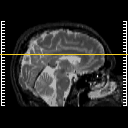
| Tour 1: Next/Previous/Start: We have moved up one slice, and the lesion we were just looking at is obscure. If you view the timeline cine, you will see it shining through from below. This gives an even better view of the edema. When contrast material is given to the patient, these regions tend to enhance, indicating that the blood-brain-barrier is disrupted. |
|
|
||||||||||||
| [Home][Help][Clinical][Tour 1][Tour 2] | Slice 33 |
| Click on sagittal image to select slice. Click on thin tickmark to change timepoint, or thick tickmark for overlay. | |
| Keith A. Johnson (keith@bwh.harvard.edu), J. Alex Becker (jabecker@mit.edu) | |

