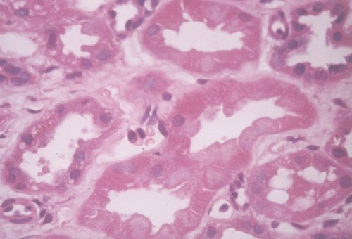
In this picture notice how the tubular epithelial cells have very eosinophilic cytoplasm and have lost their nuclei.
- The right kidney weighed 270g, the left kidney 280g.
- Capsules stripped easily to reveal a granular surface.
- Cut surfaces of both kidneys showed loss of normal corticomedullary distinction, and histology revealed that acute tubular necrosis was present in both kidneys (see picture).
- The renal pelves and ureters were normal.
- The bladder had a trabeculated appearance, and no prostate tissue could be found.