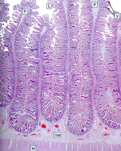
The small intestine (2)

The micrograph above shows a section of the jejunum, and clearly shows the highly folded structure of the mucosa of the small intestine. The epithelial surface of the plicae (P) is further folded to form villi(V). These increase the surface area of the small intestine still further, and the surface of each villus is covered in small microvilli to maximise surface area- the area available for absorption is vast. Each villus has its own blood supply- the vessels can be seen in the submucosa (SM)- and blood containing digestive products from the small intestine is taken to the liver via the hepatic portal system. The double muscle layer (M) moves food through the intestine by peristalisis.


