| Tour 1: Next/Previous/Start:
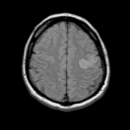
This acute cerebral infarction can be seen to involve the left pre-
central gyrus. Abnormally bright signal is seen here because of the
presence of excess water which has a prolonged relaxation time. As
tissue has become infarcted and edematous, the sulcus
is no longer identifiable. Compare the infarcted side with the normal
right side. As you navigate through the datasets, change between the
three types of image with the buttons at right, or click on the timeline
tickmark for the desired dataset.
|
|


