 |
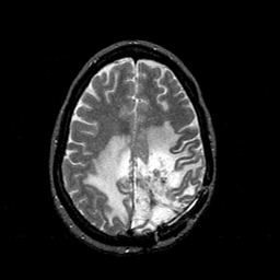
| Tour 1: Next/Previous/Start: This tour will examine the left parietal lesion, found at biopsy to be an anaplastic astrocytoma. First, note that the T2-weighted image shows a poorly circumscribed mass with a core of mixed high and low signal, a rounded periphery of higher signal, and a component of infiltrating edema, appearing as somewhat lower signal. The tumor appears to be centered in white matter underlying the left post-central gyrus. |
|
|
|||||||||||||
| [Home][Help][Clinical][Tour 1][Tour 2][Tour 3] | Slice 37 |
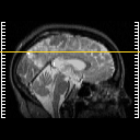
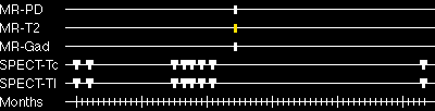
| Click on sagittal image to select slice. Click on thin tickmark to change timepoint, or thick tickmark for overlay. | |
| Keith A. Johnson (keith@bwh.harvard.edu), J. Alex Becker (jabecker@mit.edu) | |