Skin and hair coloration is determined by the pigment melanin which is produced in cells called melanocytes. The inability of these cells to produce melanin or the absent of these cells results in a lighter or completely white skin. Vitiligo is a condition where white patches, or macules, develop on the skin. It may involve both the pigmentation of the skin and hair (1). Generally it is agreed that there are no functional melanocytes or pigment cells in vitiligo skins. Even recent histochemical and immunological studies on vitiliginous skins could not detect identifiable melanocytes in the white macules (2,3). Melanocytes are normally found in the basal layer of the epidermis of the skin. Refer to MEDIC EDUCATION:Histology for more information on the anatomy of the skin and pictures of melanocytes.
Although vitiligo is not lethal, the physical disfigurement perceived by those afflicted leads to social embarrassment and psychological turmoil. Therefore, it should not be considered as a mere cosmetic disorder. See the pictures below.
)window.location='http://vflylab.calstatela.edu/edesktop/WWW_Projects/Medical/Vitiligo_esarkis/Images/2078.jpg')
)window.location='http://vflylab.calstatela.edu/edesktop/WWW_Projects/Medical/Vitiligo_esarkis/Images/1540.jpg')
)window.location='http://vflylab.calstatela.edu/edesktop/WWW_Projects/Medical/Vitiligo_esarkis/Images/1825.jpg')
Return to Table Of Content
the incidence of vitiligo is estimated to be 1% in our population. Vitiligo occurs worldwide and it affects all races Obviously it is of greater concern for people with dark skin as there will be drastic contrasts. Also, vitiligo shows no preference over any one of the sexes. There is a positive family history in about 30% of cases (4). There seems to be no preference over any specific hair or eye coloration. Vitiligo may appear from birth to senescence, but approximately one half of the patient with vitiligo acquire it before the age of 20 (1,4). See vitiligo for more general information on the disease.
Return to Table Of Content
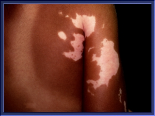
Vitiligo macules are characteristically uniformly chalk-white. On occasion the border of the depigmented area may have an intermediate tan color. The macules have fairly discrete margins and are rounded or linear in shape. Except for the change in color, vitiligo skins otherwise usually appear normal. although vitiligo may occur anywhere on the skin, there are characteristic patterns of involvement (1). )window.location='http://vflylab.calstatela.edu/edesktop/WWW_Projects/Medical/Vitiligo_esarkis/Images/2081.jpg') Very often, the white macules are localized to the sites that are normally hyperpigmented such as the face and dorsal hands, umbilicus, nipples, and sacrum. The least afflicted regions are soles and palms which in normal skin are least pigmented. In addition, with some people the macules appear symmetrical as it is shown here(1).
Very often, the white macules are localized to the sites that are normally hyperpigmented such as the face and dorsal hands, umbilicus, nipples, and sacrum. The least afflicted regions are soles and palms which in normal skin are least pigmented. In addition, with some people the macules appear symmetrical as it is shown here(1).
Return to Table Of Content
While most vitiligo patients are generally healthy, several disorders such as thyroid disorders, Addison's disease and melanoma have been associated with vitiligo.
Melanoma is malignant cancer of the pigment cells, and some people afflicted with this disease also develop vitiligo. See melanoma for more information about this disease. The picture presents a case of melanoma. )window.location='http://vflylab.calstatela.edu/edesktop/WWW_Projects/Medical/Vitiligo_esarkis/Images/i0000012.jpg') One possible explanation is that in patients with malignant melanoma the immune system generates antibodies to the surface antigen of the melanocytes. This results in the destruction of the melanocytes, resulting in the hypopigmentation of the skin (6).
One possible explanation is that in patients with malignant melanoma the immune system generates antibodies to the surface antigen of the melanocytes. This results in the destruction of the melanocytes, resulting in the hypopigmentation of the skin (6).
Return to Table Of Content
The etiology of vitiligo is unknown. There may be one cause or many causes which result in the disappearance of the melanocytes from the skin. Three major hypotheses have been proposed to explain the pathogenesis of vitiligo. According to immune hypothesis, melanocytes are ultimately destroyed by humoral or cellular immune mechanism. The self-destruct theory suggests that there is an intermediate or metabolite in the melanin synthesis that, if unchecked, leads to the destruction of the melanocytes. Finally, neural hypothesis states that vitiligo is caused by neuronal factors, such as the accumulation of neurochemical mediators (1,10). Some researchers believe that vitiligo is not caused by only one factor but rather each of the causes mentioned above may be involved in vitiligo pathogenesis in different patients (11).
Return to Table Of Content
Vitiligo is frequently associated with several autoimmune diseases such as pernicious anemia, thyroid disease, Addison's disease, and uveitis. Therefore, melanocytes are thought to be destroyed by humoral or cellular immune mechanisms. In support of this hypothesis, it has been shown that antibodies to melanocytes-associated antigens are present in common vitiligo (12). In addition, it has been shown that the level of antimelanocyte antibodies was higher in patients with active vitiligo than in patients with inactive vitiligo or normal people (13). Studies done to investigate the role of cellular immune mechanisms have been contradictory. Whereas some researchers have reported an increase in the level of T4 cells in the blood of vitiligo patients (14), some have found a lower level of T4 cells in vitiligo patients comparing to normal people (15).
This hypothesis suggests that during melanin synthesis pathway toxic intermediates are produced that if unchecked will cause the destruction of the melanocytes, resulting in vitiligo. In other words, there is control mechanism which fails to operate normally and results in the accumulation of toxic substances. It is believed that these toxic chemicals are the highly reactive radicals, mainly superoxide radicals. Superoxide radicals, if not checked, will result in the formation of hydroxyl radicals which cause serious damage to the cells (16). The anti-radical mechanisms include thioredoxin reductase, catalase, and superoxide dismutase. Superoxide dismutase is an enzyme which scavenges oxygen radicals into hydrogen peroxide which in turn is removed by catalase. Separate studies have shown that the catalase(18) and thioredoxin reductase (19) levels are lower in the skin of vitiligo patients.
Unlike the other two hypotheses, the role of the nervous system in the pathogenesis of vitiligo is not well defined, and research done in this area is very limited. Perhaps the most important clinical evidence indicating possible neuronal involvement is a form of vitiligo called segmental vitiligo. Here the white macules are formed along specific dermatomes. Each dermatome presents a portion of the skin which is served by a specific peripheral nerve(1).
Many researchers have been using various animal models, such as the pattern of pigmentation in chicken feathers, to test the above hypotheses or to develop new ones which might help to find the causes of vitiligo. See frog .
Refer to articles for a list of recent research articles on vitiligo as well as a list of research centers.
Return to Table Of Content
Currently there are three treatments that are available to vitiligo patients. Repigmentation via photosensitive compounds and exposure to UVA light. Depigmentation of the remaining pigmented skins in order to make the skin uniform in color. Finally, grafting of pigmented tissue into the vitiligo macules (10). In psoralen-photochemotherapy, psoralen, which is a photosensitive chemical, is either ingested as tablets or applied to the white patches of skin as lotion. Subsequently, the patient is exposed to UVA light.  Thus, the treatment is called PUVA therapy. If the treatment continues for a long time high percentage of patients show recovery of pigmentation. The mechanism of psoralen photoactivation melanogenesis is unclear. Since psoralen makes the eyes sensitive to light special glasses are worn during treatment as well as after treatment for several hours to prevent possible damage to the eyes. Depigmentation of the remaining pigmented skin is another way to produce uniform skin coloration. This procedure is recommended in individuals who have lost more than 50% of their skin pigmentation. This can be done by the application of certain benzene compounds to the skin (1). Finally, during the past couple of years surgical approach for the treatment of vitiligo has been developed. One method is to graft autologous skin to vitiligo macules. The other is to apply suspensions of cultured melanocytes to vitiligo macules (15). The picture shows a person undergoing UVA therapy.
Return to Table Of Content

Visit rain for a survey of weather in your area in U.S. .
The following has been added by the Electronic Desktop Project:
 Contact Us
If you are an educator who is using our NEXTSTEP or virtual applications in the classroom, we would especially like to hear from you. Let us know what you are doing and how it is working out. Continued support for this project will depend on its impact in science education.
If you are an educator who is interested in making use of our NEXTSTEP or virtual applications, please let us know how we can help.
 Return to the Electronic Desktop Project home page
 Check out the WWW Virtual Application Catalog from the EDP
 Check out the NEXTSTEP Application Catalog from the EDP
 Visit the home page for California State University, Los Angeles