Hematopathology of Lymph Nodes,
Spleen, and Other Organs
Slide 1 of 212
Image ID 4612
Keywords
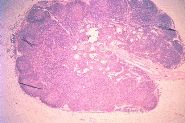
Normal lymph node
Description
An extremely low power view of an H&E stained
lymph node is seen in this photomicrograph.
Follicles are noted in the cortical area and these
are differentiated from the non-follicular portions
of the node. Vascular structures are also seen.
Click on this image
to enlarge it, then
on Back botton
in the Netscape Menu
to shrink it back down