Hematopathology of Lymph Nodes,
Spleen, and Other Organs
Slide 2 of 212
Image ID 4613
Keywords
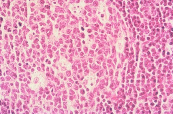
Lymph node follicle
Description
This is a low power view of a lymph node follicle
stained with H&E. The distinction between
peripheral and central areas can be readily seen.
Variation in cell type can be also seen.
Click on this image
to enlarge it, then
on Back botton
in the Netscape Menu
to shrink it back down