An Alzheimer's Primer:
Navigating through the Web
of
Plaques and Tangles


Navigating through the Web
of
Plaques and Tangles
Late onset AD can be divided into familial and nonfamilial forms. Among the two familial forms, early and late onset, late onset represents a larger proportion of cases and is associated with a locus on chromosome 14. No gene has been located yet. For research and case studies of familial Alzheimer's, refer to the Online Mendelian Inheritance in Man Database. A recently published book, Hannah's Heirs: The Quest for the Genetic Orgins of Alzheimer's Disease by Daniel A. Pollen, M.D., may also be of interest.
The majority of AD cases are late onset in which genetic involvement is unclear. However, possesing one or two copies of the E4 allele of the alipoprotein E gene on chromosome 19 is now considered a risk factor.
Stage 2: lasts 2-10 years; increasing memory loss and confusion, difficulty recognizing friends and family, perceptual and motor problems, loss of language facility and logical thinking.
Stage 3: lasts 1-3 years; inability to recognize self or family, weight loss, little or no capacity to care for self, incontinence, aphasia, difficulty swallowing, and seizures.
For a more complete list of warning signs see this list..
Neuronal Loss
Loss in neuronal numbers is a normal part of the aging process-- up to a 30% loss of neocortical neurons by age 65 occurs in healthy individuals. AD patients lose 50% or more of their neocortical neurons, primarily in the frontal and temporal lobes.
Dendritic Degeneration
The degree of degeneration of the basal dendrites of the neocortex, rather than the age of the patient, is more predictive of the extent of dementia. In presenile dementia, "lawless" dendritic growth is commonly seen. These chaotic tangles of dendrites impair normal brain functioning.
Neurofibrillary Tangles
 These are abnormally shaped neurofibrils and neurotubles that twist together in a spiraling pattern. The result is reduced flow of material in these neurons, impeding normal functioning. In fact, the density of the tangles, rather than the number of amyloid plaques (discussed below), is closely correlated with the degree of dementia. This intracellular phenomenon is present to a small degree in normal aging brains. These tangles occur in larger numbers in individuals over forty with Down Syndrome, leading researchers to look at chromosome 21 for a genetic link.
Amyloid Plaques

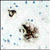 These consist of decaying nerve endings and astrocytes with a core of an amyloid beta protein fragment. This fragment comes from a glycoprotein precusor that occurs in most cells as an integral membrane protein. The amyloid protein may also be secreted from normal cells. The gene for the amyloid protein precursor is located on chromosome 21. A mutation of this gene has been implicated in early onset familial AD. To download the protein motif fingerprint of the amyloid precursors in humans, mice, and rats from the National Institutes of Health, click here. Amyloid plaques are also present in the brains of individuals with Down Syndrome. The plaques occur mainly in the cerebral cortex and hippocampus, regions which control memory, thinking, reasoning, and sensory perception --all areas of impairment in AD patients.
The question is, what causes these proteins to clump together into a plaque? Alipoprotein E is suspected of being involved in this process. One scientist's self-proclaimed "ramblings" on alipoprotein E's possible role in AD can be found here. For research on amyloid beta protein and its relationship to AD and other diseases refer to The Laboratory of Molecular Biophyisics at Oxford.
Abnormal Neurotransmitter Levels
Decreased levels of acetylcholine, norepinephrine, and serotonin are found in the brains of AD patients. Attention has focused on acetylcholine since the number of cholinergic neurons is markedly lower in AD patients. Levels of acetylcholine can be measured by the concentration of choline acetyltransferase (ChAT), the enzyme that catalyzes the synthesis of acetylcholine. ChAT levels are 60-90% lower in AD patients than in age-matched nondemented individuals. One experimental treatment is to administer drugs, such as tacrine, that will inhibit acetylcholinesterase, the enzyme that hydrolizes acetylcholine. The anticipated result is to increase levels of acetycholine since the enzyme required for its degradation is disabled. Thus far, results have not been promising. It remains to be discovered whether the decreased number of cholinergic neurons are a cause or a consequence of AD.
Although definitive diagnosis requires autopsy or biopsy, current imaging techiniques have improved the diagnostic ability to 90% accuracy.This is possible because the brains of AD patients show areas decreased blood flow and shrinkage. These changes can be seen in single photon emission computed tomography (SPECT) and magnetic resonance imaging (MRI) scans, respectively. Areas of decreased blood flow indicate areas of decreased functioning. In SPECT images, decreased blood flow is seen as blue to black, whereas regions of high blood flow are orange to white.

The following  movie is an overlay of SPECT images and MRI scans.
The Whole Brain Atlas offers tours through an Alzheimer's brain using
 MRI images or
  SPECT images. Go directly to the Whole Brain Atlas to view hundreds of MRI's of a normal brain. To learn more about imaging of the Alzheimer's brain.
 Contact Us
If you are an educator who is using our NEXTSTEP or virtual applications in the classroom, we would especially like to hear from you. Let us know what you are doing and how it is working out. Continued support for this project will depend on its impact in science education.
If you are an educator who is interested in making use of our NEXTSTEP or virtual applications, please let us know how we can help.
 Return to the Electronic Desktop Project home page
![]() Check out the WWW Virtual Application Catalog from the EDP
Check out the WWW Virtual Application Catalog from the EDP
 Check out the NEXTSTEP Application Catalog from the EDP
 Visit the home page for California State University, Los Angeles