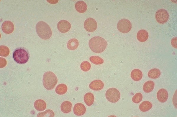
This high dry view of the Wright's stained peripheral blood
is from a patient with hereditary spherocytosis. A normal lymphocyte is
seen in the field for comparison purposes. Large, greyishred
polychromatophilic "shift" cells are seen, as are many spherocytes.
Click on this image
to enlarge it, then
on Back buttom
in the Netscape Menu
to shrink it back down